Mycorrhizae
When colonizing land habitats, the availability of sufficient water and nutrients was a challenge for plants, and mutualistic associations of thalli and roots with fungi were the most important coevolutionary processes. Arbuscular mycorrhizae (Figs. 2, 3) are known from the Ordovician (Redecker et al. 2000) and they are most common mycorrhizal partners in all groups of land plants. The evolutionary switch of certain ecologically most important plant groups to other fungi to improve symbiosis, is not yet understood. Climax vegetations in temperate zones of the Northern and partly the Southern Hemisphere are in fact obligate ectomycorrhizal communities with trees of the Pinaceae, Fagales and Salicaceae. In plant associations dominated by Ericales the ericoid mycorrhizae play an essential role. Early ontogenetic stages in orchid development depend on specific endotrophic fungal partners, and heterotrophic orchids require an obligat mycorrhization.
In Sebacinales all mycorrhizal types occur that are known in Basidiomycota. Tulasnellales appear to be frequent in orchid mycorrhizae and in Aneuraceae, but also occur in the main autotrophic partners, the Pinaceae and the Fagales.
Evolutionary trends in mycorrhizae:
Root associated fungi > hyphal sheaths > intercellular hyphal growth
Root parasites > endophytes > endomycorrhizae
Exclusively mycorrhizal: Sebacinales
Exclusively ectomycorrhizal: Thelephorales
Saprobic > ectomycorrhizal: Cantharellales, Gomphales
Wood decay > ectomycorrhizal: Russulales, Atheliales, Boletales, Agaricales
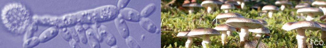
Sebacinales

Fig. 44. Sebacina epigaea in a mixed forest with fructifications on soil (arrows and insert to the right). The ectomycorrhizal species are associated with pine roots in this forest, but can grow also on roots of other ectomycorrhizal host trees. Under favorable conditions fructifications appear frequently in autumn. Orig. F. Oberwinkler.
Little attention was paid to mostly inconspicuous Sebacina species and related fungi until their obligatory association with plant roots was detected and molecular screenings revealed an enormous unknown and cryptic diversity (Weiß et al. 2011). It turned out that Sebacinales are mycobionts in ectomycorrhizae of Pinaceae, Fagales, Myrtaceae, and also in Polygonum viviparum (Mühlmann et al. 2008), living in subalpine and alpine grass vegetations. Sebacinales also constitute orchid mycobionts (Warcup 1988, Selosse et al. 2002b, Weiß et al. 2004b, Suárez et al. 2008) and they are capable to form ericoid, arbutoid, and cavendishioid mycorrhizae (Berch et al. 2002, Selosse et al. 2002a, Setaro et al. 2006a,b, Selosse et al. 2007, Kottke et al. 2008). In addition, Sebacinales associate with thalli of jungermannioid liverworts (Kottke et al. 2003, Nebel et al. 2004).
Evolutionary trends in Sebacinales:
Basidiocarps lacking > resupinate > pustulate > erumpent > stereoid > clavarioid
Hyphae thin-walled > thick-walled
Growth saprobic > endophytic > mycorrhizal
Liverwort associates > ectomycobionts of Pinaceae, Fagales and others
Liverwort associates > endomycorrhizae in orchids and Ericales
As in nearly all other monophyla of the Agaricomycotina, also in the Sebacinales basidiocarps evolved from inconspicuous generative hyphal networks to corticioid, pustulate, stalked-stereoid, and clavarioid structures (Fig. 45). More elaborate basidiomata and hymenia, like porioid, hydnoid, or agaricoid are not known. Hyphae are clampless at least in species with basidiocarps. Rarely, thick-walled hyphae occur, as in Sebacina dimitica. Basidia are longitudinally septate, and basidiospores often germinate with secondary spores.
As the Thelephorales, also Sebacinales appear to be exclusively mycorrhizal fungi. Ectomycorrhizae, arbutoid and orchid mycorrhizae have only been found in group A of a phylogenetic tree, while ericoid and cavendishioid mycorrhizae are restricted to group B (Weiß et al. 2004b, Selosse et al. 2007, Weiß et al. 2011). It is premature to interprete evolutionary trends in these clusterings. Also, host specificities cannot be recognized so far. As in other mycorrhizal taxa, major evolutionary steps in land plants, liverworts, Pinaceae, Fagales, Ericales, orchids and others, certainly had a strong influence on adaptive radiations of Sebacinales, but these are not yet understood.

Fig. 45. Fructifications of Sebacinales and micromorphology of Sebacina incrustans. Sebacina species produce resupinate and incrusting basidiocarps on soil, litter and wood on the forest floor. Efibulobasidium and Craterocolla have gelatinous fructifications growing on dead wood. Tremellodendron and Tremelloscypha species grow on the forest floor. The micromorphology of Sebacina incrustans is typical also for other Sebacinales. Orig. F. Oberwinkler.
Tulasnellales
Because of the unique basidial development and morphology (Fig. 46) and their importance as liverwort and orchid mycorrhizal fungi, it seems appropriate to keep Tulasnellales as an order separate from the Cantharellales.

Fig. 46. Tulasnella spp. a Fully developped basidiocarp of Tulasnella sp. on the underside of a Betula pubescens-trunk. b Hymenium of Tulasnella calospora with different stages of basidial development. c SEM photograph of Tulasnella vermispora, one spore attached to the sterigma. Orig. F. Oberwinkler.
Evolutionary trends in Tulasnellales:
Basidiocarps lacking > resupinate
Hyphae with clamps > unclamped
Growth parasitic? > endophytic > mycorrhizal
Mycothalli > endomycorrhizae in orchids > ectomycorrhizae of Pinaceae, Fagales and others
The thallose species of Aneuraceae, a family of the Metzgeriales, have Tulasnella mycobionts (Nebel et al. 2004, Preussing et al. 2010) and were considered by Krause et al. (2011) as a model of early evolved symbiotic associations. Cryptothallus mirabilis is a myco-heterotrophic liverwort and specialized as an epiparasite on Tulasnella species that form ectomycorrhizae with surrounding trees like Betula pubescens, Pinus pinaster and P. muricata (Bidartondo et al. 2003). – Tulasnella spp. as mycobionts in orchids have been reported from various parts of the world (e.g. Shefferson et al. 2005, 2007, Suárez et al. 2009, Cruz et al. 2010, Yuan et al. 2010). Even when Tulasnellales are the preferred mycobionts of orchids, Sebacinales, Thelephorales, Agaricales, and also Tuberales associate with them.
In a molecular screening of orchid mycorrhizae from Southern Ecuador (Kottke et al. 2009), sequence taxa clustered with the Atractiellomycetes, a relationship of the Pucciniomycotina. A specific cell organelle, the symplechosome (Fig. 16), found in intracellular hyphae, confirmed the molecular identification of these fungi. So far, this finding is unique and requires confirmation trough additional sampling. The origin of these fungi remains unclear. There are no other mycorrhizal fungi known in the Pucciniomycotina and also not in the Ustilaginomycotina.
Cantharellales
The cantharelloid clade, as circumscribed in phylogenetic hypotheses by Moncalvo et al. (2006) comprises the genera Botryobasidium, Sistotrema, Clavulina, Multiclavula, Craterellus, Cantharellus, and Hydnum. The authors also included the Ceratobasidiaceae and Tulasnellaceae in the Cantharellales. Species of Botryobasidium and Sistotrema are saprotrophs, Multiclavula species are basidiolichens, and Clavulina, Craterellus, Cantharellus, and Hydnum are ectomycorrhizal fungi. There is no synapomorphy known for the taxa included in the Cantharellales.

Fig. 47. Morphology of Cantharellus cibarius. a-d basidiocarps in different developmental stages, c longitudinal section. e hyphal arrangement of pileus surface, f subhymenial hyphae, g part of the hymenium with basidia of different ages, h basidiospores. After Oberwinkler (1977), modified with additions.
Evolutionary trends in Cantharellales:
Basidiocarps resupinate > clavarioid > stalked capitate
Hymenium smooth > irregular > hydnoid > cantharelloid
Saprotrophic > lichenized
Saprotrophic > ectomycorrhizal
The evolutionary transitions from crustose to cantharelloid fructifications cannot be reconstructed. Also the origin of lichenization remains unclear, but all Multiclavula species are clavarioid. According to the proposed phylogenetic hypothesis of Moncalvo et al. (2006), Cantharellus and related genera constitute a clade separate from Clavulina, thus indicating that ectomycorrhizal fungi evolved at least twice in Cantharellales.
Thelephorales
Most species of the Thelephorales have brownish pigmented and characteristicly ornamented basidiospores (Fig. 48). Thelephoric acid is common and all species analyzed so far are mycobionts in mycorrhizae of seed plants. Basidiocarps and hymenia display a convergent series of corticioid, ondontioid, lenzitoid, thelephoroid-clavarioid, hydnoid and boletoid structures, but agaricoid and gasteroid basidiomata are not known (Fig. 11). All relevant phylogenetic hypotheses, based on molecular data, confirm the monophyly of the Thelephorales.
Evolutionary trends in Thelephorales:
Basidiocarps resupinate > stereoid > clavarioid > stalked capitate
Hymenium smooth > irregular > hydnoid > cantharelloid
Origin unknown > mycorrhizal
Many species of the Tomentella-Thelephora relationship and those of the Bankeraceae grow on soil in forests. In addition, many Tomentella species produce basidiocarps on wood and were therefore formerly considered a saprotrophs. However, all molecularly analyzed species could be identified as mycorrhizal partners (e.g. Bruns et al. 1998, Kõljalg et al. 2000, 2001, 2002). Thus, it is very likely that all Thelephorales are mycobionts with unknown origin. Also the distribution patterns with their hosts cannot be explained along evolutionary trends.

Fig. 48. Spore morphology, basidiocarps and hymenial configurations in Thelephorales. Typical basidiospores of the Thelephoraceae have pigmented and tubercular walls with spiny protuberances. Resupinate Tomentella species are very common in forests with acidic soils. Sometimes, basidiocarps of Thelephora begin to grow corticioid and continue irregularly stereoid-thelephoroid. Stalked capitate basidicarps with hydnoid hymenia are typical for the Bankeraceae. Boletopsis is an imitation of Boletus, but shares the micromorphology of the Thelephorales. Orig. F. Oberwinkler.
Russulales
Amyloid spore ornamentation together with gloeoplerous hyphae constitute a set of synapomorphies that characterize species of the Russulales (Fig. 49). It was provocative to postulate a relationship in „premolecular times“ that circumscribed basidiomycetous fungi with resupinate to gasteroid basidiomata, including nearly all other fruting body structures, and various hymenophore configurations (Oberwinkler 1977).

Fig. 49. Micromorphology, basidiocarps and main trophic stages in Russulales. The hymenial detail of Bondarzewia montana shows also the characteristic feature of gloeoplerous hyphae, the warty spore ornaments are amyloid. Some of the most characteristic basidiocarps are illustrated by representative genera. The distribution of white-rot and ectomycorrhizal fungi in the scheme applies for the Russulaceae. Orig. F. Oberwinkler.
Evolutionary trends in Russulales:
Basidiocarps resupinate > stereoid > discoid > clavarioid > pileate > gasteroid
Hymenium smooth > irregular > hydnoid > porioid > lamellate
Saprotrophic > parasitic
White rot > brown rot
Saprotrophic > ectomycorrhizal
Based on molecular and morphological data, Miller et al. (2006) recognized 12 families and approximately 80 genera in the Russulales. Only Albatrellaceae and Russulaceae contain ectomycorrhizal taxa and represent separate clades. Albatrellacease comprise predominantly pileate poroid species, however Byssoporia terrestris is resupinate and Leucogaster, Leucophlebs and Mycolevis are gasteroid. In Russulaceae Lactarius and Russula contain many species with a global distribution in ectomycorrhizal vegetations, in temperate regions very frequently with dominating Pinaceae and Fagales. Coevolutionary processes reached species-species dependencies in many cases (Fig. 50). Basidiocarps in Russulaceae are exclusively agaricoid and gasteroid, the latter e.g. Arcangeliella, Cystangium, Gymnomyces, Macowanites, Martellia, and Zelleromyces. Most of these gasteroid genera appear to be paraphyletic. – The switches from white rotting ancestors to brown rot decay fungi and to ectomycorrhizal ones are unresolved.

Fig. 50. Host dependencies in Lactarius sect. Dapetes. Lactarius deterrimus is restricted to Picea abies, L. salmonicolor to Abies alba. Lactarius deliciosus, L. sanguifluus, and L. semisanguifluus are associated with Pinus sylvestris, but prefer different soil conditions as indicated in die diagram. Orig. F. Oberwinkler.
Atheliales
The Atheliales is composed of resupinate species with loose subhymenia, smooth to slightly irregular hymenia (Fig. 51), and an unusual diversity of trophic stages (Fig. 17), including saprotrophs, algal and lichen parasites as well as animal and plant symbionts (Binder et al. 2005). The lichenized Lepidostromataceae (Ertz et al. 2008) appears to be the sister clade.
Evolutionary trends in Atheliales:
Cystidia lacking > present
Basidiospores smooth > lobate > bluntly warty
Saprotrophs > parasites > symbionts
Saprotrophic > ectomycorrhizal
Most of the major clades in the Agaricomycotina have corticioid species in phylogenetically basal positions, thus confirming convergent evolutionary trends from simple to complex ones (Fig. 11). In the Atheliales, however, such an evolution of basidiomata is lacking. Micromorphological features, like hymenial cystidia in Amphinema byssoides, or oramented spores, as in species of the genus Tylospora (Fig. 51) cannot be interpreted in an evolutionary context. Also, the diversity of trophic stages is enigmatic, even when species of Amphinema, Byssocorticium, Piloderma and Tylospora are important and widespread ectomycorrhizal mycobionts.

Fig. 51. Basidiocarps of representative members of the Atheliales. a Athelia epiphylla, b Tylospora asterophora and c Amphinema byssoides. Each of the figures illustrates the complete cellular construction of the species, except the hyphae in the substrate. Characteristic are the loose subhymenia and the non-thickening hymenia. a from Oberwinkler (1977), orig. F. Oberwinkler.
Boletales
Fusiform, thick-walled and strongly pigmented basidiospores are the most common ones in Boletales. In frequent cases the hyphal system is monomitic and soft, and the large number of pigments are derivatives of pulvinic acid. These features were applied to circumscribe the Boletales in premolecular times. All phylogenetic hypotheses, based on molecular data, support the order. Basidiocarps range from resupinate to gasteroid with a major radiation in bolets (Figs. 11, 52).
Evolutionary trends in Boletales:
Basidiocarps resupinate > merulioid > porioid > pileate > gasteroid
Hymenium smooth > merulioid > hydnoid > poroid > boletoid > lamellate
Basidiospores smooth-walled > reticulate
brown rot > ectomycorrhizal > mycoparasitic
As in the Atheliales, also in Boletales resupinate basidiocarps and brown rot saprotrophism could be ancestral states (Binder & Hibbett 2006). White rot is not known in these fungi, but a specific mode of brown-rot was developed by Coniophoraceae on conifers. Ectomycorrhizal associations are known from Pinaceae, Fagales, Fabales, Myrtaceae, Salicaceae and the tropical Dipterocarpaceae, and ericoid mycorrhizae with Ericales. A switch from brown rotting Serpula to ectomycorrhizal Austropaxillus and Gymnopaxillus species in Nothofagus and Eucalyptus forests has been reported by Claridge et al. (2001). Mycoparasitism of Pseudoboletus parasiticus on Scleroderma citrinum is known for a long time, but parasitic interactions of Chroogomphus and Gomphidius spp. on ectomycorrhizae of Suillus and Rhizopogon spp. have been discovered recently (Agerer 1987-1998). Ectomycorrhizal capabilities of Pisolithus parasiticus appear as not efficient enough for the required nutrient supply (Raidl 1997). Reductions and losses in specific protein families were found in functional genomics of Serpula lacrymans (Eastwood et al. 2011), and interpreted as adaptations to intercellular interactions with plant tissues. The known host specificity of Leccinum spp. could be confirmed and reconstructed using a molecular clock by den Bakker et al. (2004). However, Leccinum aurantiacum has a broad range of host trees.

Fig. 52. Basidiocarps and hymenial micromorphology in Boletales. Part of the hymenium and subhymenium of Tylopilus felleus shows characteristic hyphal arrangement, basidial and spore morphology in Boletales. Basidiocarp illustrations of representative genera are arranged from resupinate to merulioid, porioid, agaricoid, boletoid, and gasteroid. Brown rot and ectomycorrhizae are the main trophic stages in Boletales. Orig. F. Oberwinkler.
Agaricales
Molecular phylogenies of the Agaricomycetes and Agaricales, based on comprehensive samplings (e.g. Matheny et al. 2006, Garnica et al. 2007, Binder et al. 2010) provided evidences for monophylies in these taxa and their subgroups. Homoplasies appear to be frequent in Agaricales, e.g. the multiple convergent evolution of sequestrate and non-gilled taxa. Morphological and/or ecological synapomorphies are not known in the order. However, several evolutionary trends may provide relevant information.
Evolutionary trends in Agaricales:
Basidiocarps clavarioid > agaricoid
Basidiocarps agaricoid > cyphelloid
Basidiocarps agaricoid > sequestrate
Basidiospores smooth-walled > ornamented
Basidiospores hyaline > pigmented
Basidiospores thin-walled > thick-walled > with germ pore
saprobic > ectomycorrhizal
saprobic > mycoparasitic
saprobic > lichenized
In several phylograms, clavarioid species, assigned to different genera, occur in basal positions of the Agaricales (Matheny et al. 2006, Garnica et al. 2007, Binder et al. 2010). Considering non-agaricoid relationships, Larsson et al. (2004) found that Typhula and Macrotyphula are closely related to some corticioid fungi, including Coronicium. – The reduction of agaricoid basidiocarps to non-lamellate ones happened at least 12 times in the Agaricales (Bodensteiner et al. 2004). Major clades comprise Schizophyllum with Fistulina and Porodisculus, Calyptella and Stigmatolemma, Cyphellopsis with Merismodes, Calathella, Lachnella and the marine Halocyphina and Nia. Surprising is the split of Hennigsomyces with Rectipilus in two separate clades. As mentioned above, cyphellization is not restricted to Agaricales. – Convergent gasteromycetation is a widespread evolutionary mode amongst Basidiomycota and occurred exceptionally frequent in the Agaricomycetes. There are sequestrate relatives inter alia in Amanita, Laccaria, Cortinarius, Coprinus, and Hebeloma. Nidulariaceae are sister to Cystoderma, and Lyoperdaceae to Agaricaceae. The merulioid Lindtneria trachyspora and the hypogeous gasteromycete Stephanospora caroticolor share a similar micromorphology, especially in basidiospore characters. To accomodate these species, Oberwinkler & Horak (1979) erected the Stephanosporace that was confirmed in a molecular phylogeny by Larsson (2007) who included also the corticioid Cristinia helvetica and Athelidium aurantiacum. – A general evolutionary trend in basidiospore morphology is an increasing complexity of the spore wall. Thick-walled, ornamented and strongly pigmented basidiospores are typical for derived Agaricales (Garnica et al. 2007). Such specialized propagules are well adapted to stressful environmental conditions.
Saprotrophic and symbiotic nutrition modes are the common ones in Agaricales, wood-decay, mycoparasitism and lichenization are comparatively rare. Main ectomycorrhizal genera include the convergently evolved Hygrophorus, Amanita, Tricholoma, Laccaria, Cortinarius, Inocybe, Hebeloma and their sequestrate relatives. Cortinarius (Fig. 53) has a worldwide distribution in ectotrophic forests and comprises at least 2000 species. Martin et al. (2008) suggested that the ectomycorrhizae-specific small secreted proteins of Laccaria bicolor have a decisive role in the establishment of the symbiosis, and that the availability of the genome will provide further insights in the functional aspects of nutrient transfers in forest ecosystems. Matheny et al. (2009) assumed that the Inocybaceae diversified no later than the Cretaceous in association with angiosperms in the Palaeotropics and had transitions to conifers possibly in the Paleogene. – Mycoparasites have been discussed above and basidiolichens will be considered in the following part.

Fig. 53. Cortinarius violaceus, the type species of the genus. a basidiocarp, b detail of subhymenium and hymenium with basidia and cystidium, c hyphal context of pileus surface, d, e basidiospores, f TEM picture of the basidiospore wall. Orig. F. Oberwinkler.